
Copy of Slicing Protocol
Preparation:
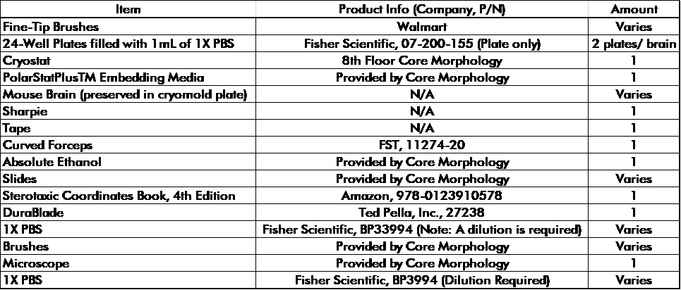
Materials
Proper Orientation of the Brain
The brain should always be positioned with the posterior side facing you. Ensure the superior side of the brain is right side up. This orientation is identical to the embedding protocol. The orientation should be followed when placing it on the circular accessory/brain holder of the cryostat.
What is the cryostat and how to use it?
The cryostat is a machine used to slice brains at a certain thickness. For our purposes, we set the cryostat to slice at a thickness of 50 microns. The cryostat is composed of the following components: side rotor, side-to-side panel, glass cover, brain holder, window, side panel (left side of cryostat), overview panel and circular accessory.
The side rotor moves the brain holder in a circular motion (slices the brain). When slicing the brain, a fluid, swift motion should be used to get the best possible section. The side-to- side panel is located within the cryostat. It holds the glass cover and blade. It has three levers that lock it in place. These levers, when loosened, adjust the angle, horizontal direction, and holder of the blade. DO NOT move the lever that controls the angle of the side-to-side panel. The glass cover is attached to the side-to-side panel. It is rotated onto the blade portion of the side-to-side panel when you want to collect sections. The brain holder is located within the cryostat. It is compromised of a screw and a hole, which holds the circular accessory. The window separates the outside environment from the cryostat’s internal environment. The optimal temperature we want the cryostat to operate at is -20°C. If the temperature deviates in any fashion, it will cause tears within the tissue. Thus, keeping the window closed as often as possible is of the utmost importance. The side panel is located left of the cryostat. It contains settings to select the section thickness and to move the brain holder forward or reverse. To adjust the section thickness, press the trim section button and use the “+” or “-“ button to either increase or decrease the thickness. Press the trim section button again to lock in the selected value. Ensure the side panel and top panel agree with the section thickness. The arrows near the bottom of the panel move the brain holder at varying speeds (Adjust as needed). The overview panel controls the light source, temperature and locks/unlocks the equipment. To lock/unlock the cryostat, hold the key button down for a few seconds. This step must be done first before anything else can be used. The circular accessory is located within the cryostat. It serves as a platform to hold the brain. When it is prepped as detailed in the procedure, place it in the brain holder and tighten the screw.
How to Use the Stereotaxic Coordinates Book
The Stereotaxic Coordinates Book provides images of sections of a mouse brain. This book serves as a map to determine where you are within the mouse’s brain as you slice and collect tissues. Use landmarks such as ventricles and markings to help you identify where you are. Since we slice the brain from posterior to anterior, begin by looking at the images from the back of the book.
How to sign up to use the cryostat
The cryostat is located on the 8th floor of the medical school building. The room is labeled as the “Core Morphology” room. To sign up, enter the room, take a right, and reserve a time on the calendar that is taped to the wall. Slicing one brain generally takes 2 hours.
Procedure:

